References

Guo S, DiPietro LA. Factors affecting wound healing. J Dent Res. 2010; 89:(3)219-229 https://doi.org/10.1177/0022034509359125

Perez Gutierrez RM, Vargas S R. Evaluation of the wound healing properties of Acalypha langiana in diabetic rats. Fitoterapia. 2006; 77:(4)286-289 https://doi.org/10.1016/j.fitote.2006.03.011

Buganza Tepole A, Kuhl E. Systems-based approaches toward wound healing. Pediatr Res. 2013; 73:(4-2)553-563 https://doi.org/10.1038/pr.2013.3

Falanga V. Wound healing and its impairment in the diabetic foot. Lancet. 2005; 366:(9498)1736-1743 https://doi.org/10.1016/S0140-6736(05)67700-8

Toker S, Gulcan E, Çaycı MK Topical atorvastatin in the treatment of diabetic wounds. Am J Med Sci. 2009; 338:(3)201-204 https://doi.org/10.1097/MAJ.0b013e3181aaf209

Mantle D, Gok MA, Lennard TW. Adverse and beneficial effects of plant extracts on skin and skin disorders. Adverse Drug React Toxicol Rev. 2001; 20:(2)89-103

Nayak BS, Isitor G, Davis EM, Pillai GK. The evidence based wound healing activity ofLawsonia inermis Linn. Phytother Res. 2007; 21:(9)827-831 https://doi.org/10.1002/ptr.2181

Tong M, Tuk B, Hekking IM, Pleumeekers MM, Boldewijn MB, Hovius SE Heparan sulfate glycosaminoglycan mimetic improves pressure ulcer healing in a rat model of cutaneous ischemia-reperfusion injury. Wound Repair Regen. 2011; 19:(4)505-514 https://doi.org/10.1111/j.1524-475X.2011.00704.x

Oryan A, Naeini AT, Moshiri A, Mohammadalipour A, Tabandeh MR. Modulation of cutaneous wound healing by silymarin in rats. J Wound Care. 2012; 21:(9)457-464 https://doi.org/10.12968/jowc.2012.21.9.457

Hashemnia M, Javdani M, Nikousefat Z Evaluation of the wound healing activity of methanolic extract of Tragopogon porrifolius in rat. Res Opin Anim Vet Sci. 2014; 4:(8)446-452

Pourgholami MH, Majzoob S, Javadi M, Kamalinejad M, Fanaee GH, Sayyah M. The fruit essential oil of Pimpinella anisum exerts anticonvulsant effects in mice. J Ethnopharmacol. 1999; 66:(2)211-215 https://doi.org/10.1016/S0378-8741(98)00161-5

Kosalec I, Pepeljnjak S, Kustrak D. Antifungal activity of fluid extract and essential oil from anise fruits (Pimpinella anisum L., Apiaceae). Acta Pharm. 2005; 55:(4)377-385

Özcan MM, Chalchat JC. Chemical composition and antifungal effect of anise (Pimpinella anisum L.) fruit oil at ripening stage. Ann Microbiol. 2006; 56:(4)353-358 https://doi.org/10.1007/BF03175031

Ghoshegir SA, Mazaheri M, Ghannadi A, Feizi A, Babaeian M, Tanhaee M Pimpinella anisum in the treatment of functional dyspepsia: A double-blind, randomized clinical trial. J Res Med Sci. 2015; 20:(1)13-21

Shojaii A, Abdollahi Fard M. Review of Pharmacological Properties and Chemical Constituents of Pimpinella anisum. ISRN Pharm. 2012; 2012:1-8 https://doi.org/10.5402/2012/510795

Jamshidzadeh A, Heidari R, Razmjou M, Karimi F, Moein MR, Farshad O An in vivo and in vitro investigation on hepatoprotective effects of Pimpinella anisum seed essential oil and extracts against carbon tetrachloride-induced toxicity. Iran J Basic Med Sci. 2015; 18:(2)205-211 https://doi.org/10.22038/IJBMS.2015.4026

Tabanca N, Ma G, Pasco DS, Bedir E, Kirimer N, Baser KH Effect of essential oils and isolated compounds from Pimpinella species on NF-κB: a target for antiinflammatory therapy. Phytother Res. 2007; 21:(8)741-745 https://doi.org/10.1002/ptr.2154

Rahimi R, Asadollahpoor A, Abdollahi M. Pimpinella anisum L. fruit: Chemical composition and effect on rat model of nonalcoholic fatty liver disease. J Res Med Sci. 2017; 22:(1) https://doi.org/10.4103/1735-1995.202147

Rajeshwari U, Shobha I, Andallu B. Comparison of aniseeds and coriander seeds for antidiabetic, hypolipidemic and antioxidant activities. Spatula DD - Peer Reviewed Journal on Complementary Medicine and Drug Discovery. 2011; 1:(1)9-16 https://doi.org/10.5455/spatula.20110106123144

Al Mofleh IA, Alhaider AA, Mossa JS, Al-Soohaibani MO, Rafatullah S. Aqueous suspension of anise “ Pimpinella anisum ” protects rats against chemically induced gastric ulcers. World J Gastroenterol. 2007; 13:(7)1112-1118 https://doi.org/10.3748/wjg.v13.i7.1112

Malveira Cavalcanti J, Henrique Leal-Cardoso J, Leite Diniz LR, Gomes Portella V, Oliveira Costa C, Barreto Medeiros Linard CF The essential oil of Croton zehntneri and trans-anethole improves cutaneous wound healing. J Ethnopharmacol. 2012; 144:(2)240-247 https://doi.org/10.1016/j.jep.2012.08.030

Kumar MS, Kirubanandan S, Sripriya R, Sehgal PK. Triphala promotes healing of infected full-thickness dermal wound. J Surg Res. 2008; 144:(1)94-101 https://doi.org/10.1016/j.jss.2007.02.049

Oryan A, Mohammadalipour A, Moshiri A, Tabandeh MR. Avocado/soybean unsaponifiables: a novel regulator of cutaneous wound healing, modelling and remodelling. Int Wound J. 2015; 12:(6)674-685 https://doi.org/10.1111/iwj.12196

Neuman RE, Logan MA. The determination of collagen and elastin in tissues. J Biol Chem. 1950; 186:(2)549-556

Mohammadalipour A, Karimi J, Khodadadi I, Solgi G, Hashemnia M, Sheikh N Dasatinib prevent hepatic fibrosis induced by carbon tetrachloride (CCl 4) via anti-inflammatory and antioxidant mechanism. Immunopharmacol Immunotoxicol. 2017; 39:(1)19-27 https://doi.org/10.1080/08923973.2016.1263860

Kalantar M, Kalantar H, Houshmand G Protective effect of hydroalcoholic extract of Lavandula officinalis l. on gentamicin induced nephrotoxicity in rats. J Babol Univers Med Sci. 2016; 18:(7)62-67

Bahabadi M, Mohammadalipour A, Karimi J Hepatoprotective effect of parthenolide in rat model of nonalcoholic fatty liver disease. Immunopharmacol Immunotoxicol. 2017; 39:(4)233-242 https://doi.org/10.1080/08923973.2017.1327965

Tellechea A, Kafanas A, Leal EC Increased skin inflammation and blood vessel density in human and experimental diabetes. Int J Low Extrem Wounds. 2013; 12:(1)4-11 https://doi.org/10.1177/1534734612474303

Gülçın İ, Oktay M, Kıreçcı E, Küfrevıoǧlu Öİ. Screening of antioxidant and antimicrobial activities of anise (Pimpinella anisum L.) seed extracts. Food Chem. 2003; 83:(3)371-382 https://doi.org/10.1016/S0308-8146(03)00098-0

Shobha R, Rajeshwari C, Andallu B. Anti-peroxidative and anti-diabetic activities of aniseeds (Pimpinella anisum L.) and identification of bioactive compounds. Am J Phytom Clin Therap. 2013; 1:516-527

Kumari M, Br E, Amberkar M Wound healing activity of aqueous extract of Crotalaria verrucosa in Wistar albino rats. Asian Pac J Trop Med. 2010; 3:(10)783-787 https://doi.org/10.1016/S1995-7645(10)60187-3

Nayak BS, Pinto Pereira LM. Catharanthus roseus flower extract has wound-healing activity in Sprague Dawley rats. BMC Complement Altern Med. 2006; 6:(1) https://doi.org/10.1186/1472-6882-6-41

Hu SC, Lan CC. High-glucose environment disturbs the physiologic functions of keratinocytes: focusing on diabetic wound healing. J Dermatol Sci. 2016; 84:(2)121-127 https://doi.org/10.1016/j.jdermsci.2016.07.008

Kunkemoeller B, Kyriakides TR. Redox signaling in diabetic wound healing regulates extracellular matrix deposition. Antioxid Redox Signal. 2017; 27:(12)823-838 https://doi.org/10.1089/ars.2017.7263

Changizi-Ashtiyani S, Seddigh A, Najafi H Pimpinella anisum L. ethanolic extract ameliorates the gentamicin-induced nephrotoxicity in rats. Nephrology (Carlton). 2017; 22:(2)133-138 https://doi.org/10.1111/nep.12953

Courtois MC, Sapoval M, Del Giudice C [Distal revascularization in diabetic patients with chronic limb ischemia]. J Mal Vasc. 2015; 40:(1)24-36 https://doi.org/10.1016/j.jmv.2014.12.007

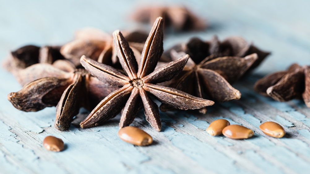
Wound healing activity of Pimpinella anisum methanolic extract in streptozotocin-induced diabetic rats
01 October 2019

Abstract

Objective:

To assess the wound healing potential of Pimpinella anisum on cutaneous wounds in diabetic rats.

Method:

Full-thickness excisional wounds were made on the back of male, Sprague-Dawley rats with diabetes. The rats were randomly allocated into four treatment groups: 1ml basal cream; tetracycline (3%); Pimpinella anisum 10% for 14 days; and a control group. At days seven, 14 and 21 post-injury, five animals of each group were euthanised, and wounds were assessed through gross, histopathological and oxidant/antioxidant evaluations. Additionally, the dry matter and hydroxyproline contents of the skin samples were measured.

Results:

A total of 60 rats were used in the study. A significant decrease in the wound size was observed in treated animals with Pimpinella anisum compared with other groups during the experiment. Additionally, treatment with Pimpinella anisum decreased the number of lymphocytes and improved the number of fibroblasts at the earlier stages and increased a number of fibrocytes at the later stages of wound healing. Other parameters such as re-epithelialisation, tissue alignment, greater maturity of collagen fibres and large capillary-sized blood vessels revealed significant changes when compared with the control. Pimpinella anisum significantly reverted oxidative changes of total antioxidant capacity, malondialdehyde and glutathione peroxidase induced by diabetic wounds (p<0.05). Furthermore, it significantly increased the dry matter and hydroxyproline contents at various stages of wound healing (p<0.05).

Conclusion:

The present study showed that application of Pimpinella anisum extract promotes wound healing activity in diabetic rats. The wound-healing property of Pimpinella anisum can be attributed to the phytoconstituents present in the plant.

Wound healing is a normal biological process of repair, following injury to the skin and other soft tissues, which is achieved through overlapping phases such as haemostasis, inflammation, proliferation and maturation.1 A fibrous scar containing large amounts of collagen is an end product of this process. Well-vascularised granulation tissue that is made within the wound area is responsible for synthesis of collagen, and provides strength and integrity to the dermis.2,3 Many important factors such as malnutrition, ageing, medication, radiation, and some diseases including diabetes, hypertension and obesity are associated with delayed wound healing.1,4

Wounds in patients with diabetes can be hard-to-heal, taking months despite essential and appropriate care.4,5 However, the exact pathogenesis of poor wound healing in patients with diabetes is not completely clear. The decrease of collagen content due to reduced biosynthesis and/or accelerated degradation of newly synthesised collagen and also the oxidative and inflammatory changes are thought to be the main causes.2,5

Register now to continue reading

Thank you for visiting Journal of Wound Care's Silk Road Supplement and reading some of our peer-reviewed resources for healthcare professionals across Asia. To read more, please register today.